| The stigma associated with brain monitoring equipment is gradually being lifted as people begin to understand how biofeedback can be used to improve mental stamina in athletes, artists and as an aid in gradually changing established patterns of behavior, attention, arousal and performance that are created and maintained at deep neurophysiological levels.
Neurofeedback is a learning strategy that enables people to alter their brainwaves and exercise the brain's own regulatory mechanisms. Also known as EEG biofeedback, it is used for many conditions and disabilities in which the brain is not functioning as well as it might. These patterns can be modified using brainwave training, also known as EEG Biofeedback or Neurofeedback.
Brain Scan Technology
The following are the various types of brain scan systems being used today which have accelerated our understanding of the brain.
|

|
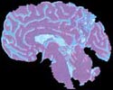
Magnetic Resonance Imaging (MRI)
Aligns atomic particles in the body tissues by magnetism, then bombards them with radio waves. This causes the particles to give off radio signals that differ according to what sort of tissue is present. A sophisticated software system called Computerized Tomography (CT) converts this information into a three-dimensional picture of any part of the body. A brain scan taken this way looks like a grayish X-ray, the different, clearly delineated types of tissue.
|

|
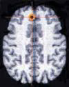
Functional MRI (fMRI)
Adds to the basic MRI anatomical picture by adding to it the areas of greatest activity. Neuronal firing is fueled by glucose and oxygen, which are carried in blood. When an area of the brain is fired up, these substances flow towards it, and fMRI shows up the areas where there is most oxygen. The latest scanners can produce four images every second. The brain takes about half a second to react to a stimulus, so this rapid scanning technique can clearly show the ebb and flow of activity in different parts of the brain as it reacts to various stimuli or undertakes different tasks. fMRI is proving to be the most rewarding of scanning techniques, but it is phenomenally expensive and brain mappers often have to share a machine with clinicians who have more pressing claims to it.
|

|
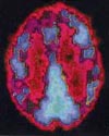
Positron Emission Topography (PET)
This technique achieves a similar end result to fMRI -- it identifies the brain areas that are working hardest by measuring their fuel intake. The pictures produced by PET are very clear (and strikingly pretty) but they cannot achieve the same fine resolution as fMRI. The technique also has a serious drawback in that it requires an injection into the bloodstream of a radioactive marker. The dose is tiny but, for safety, no one person is generally allowed to have more than 12 scans per year (this actually equals one scanning session).
|
|
Photo Not Shown
|
Electroencephalography (EEG)
Measures brainwaves - the electrical patterns created by the rhythmic oscillations of neurons. These waves show characteristic changes according to the type of brain activity that is going on. EEG measures these waves by picking up signals via electrodes placed in the skull. The latest version of EEG takes readings from dozens of different spots and compares them, building up a picture of varying activity across the brain. Brain mapping with EEG often uses Event-Related Potentials (ERPs), which simply means that an electrical peak (potential) is related to a particular stimulus like a word or a touch.
|

|
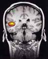
Magnetoencephalography (MEG)
Similar to EEG in that it picks up signals from neuronal oscillation, but it does it by homing in on the tiny magnetic pulse they give off rather than the electric field. This technique has weaknesses -- the signals are usually weak and there is interference. Yet, it has enormous potential because it is faster than other scanning techniques and can therefore chart changes in brain activity more accurately than fMRI or PET.
|

Descriptions & Scan Photos from:
Mapping the Brain, Rita Carter
Weidenfeld & Nicolson
|


